Oral Cancer Screening Device

OralScan

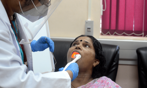
OralScan is an optical imaging multimodal device for the early detection of (pre-) cancerous lesions of the oral cavity. The device operates on the principle of diffuse reflectance and tissue autofluorescence wherein light undergoes multiple absorption and scattering events before emerging from the tissue surface. Tumour tissues undergo biochemical and morphological changes during the process of carcinogenesis, which is reflected in the optical signals emanating from the tissue. The device uses an optical system with custom-built software and algorithms for tissue analysis. OralScan enables the physician to visualize and discriminate between healthy and potentially malignant sites of the oral cavity before they perform a biopsy. The diagnostic advantages of OralScan include non-invasive in vivo procedures and services, wide-field imaging of the oral cavity, utilization of a cloud-based machine learning algorithm for real-time feedback on tissue status, and application of oxygenated haemoglobin (HbO2) absorption maps for biopsy guidance.

OralScan is a CE-marked product

Specifications :

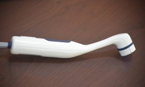
  • Non-invasive, hand-held intraoral multimodal imaging device integrated with a high resolution (5MPx) monochrome CMOS sensor (2592×1944 resolution, 2.2 micron size pixels)
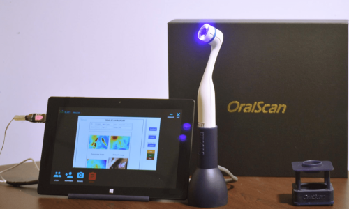
  • Tissue illumination with violet (405nm), green (542nm) and red (610nm) LEDs
  • Quantitative and real time feedback on tissue status with the help of a cloud based Machine Learning (ML) algorithm
  • Clinically validated device for screening and early detection of oral potentially malignant lesions (OPML)
  • Ideally suited for biopsy guidance
  • Portable for use along with a Windows Tablet or Laptop in remote locations
  • Quick and pain free screening
  • Trained health workers and paramedical staff can operate the instrument
  • Suitable for periodic reviews following surgical/radiation treatments

Technology

Photonics is the science and technology of generation, manipulation, and detection of photons, which is the quantum unit of light. Biophotonics is the science of intertwining light with medicine to image, detect, and manipulate biological materials based on the principles of biology, photonics, and spectroscopy. We at Sascan use light to screen tissue abnormalities at the biochemical, structural, and morphological levels and detect early signs of malignancy.  Unlike conventional techniques used for cancer detection, our technology enables multimodal imaging of tissue in a multispectral imaging platform for improved accuracy. It provides screening results immediately after the scan in real-time with the help of a cloud-based machine learning algorithm. If the tissue status warrants a biopsy, our proprietary software would process the captured multispectral images and assist the clinician for locating the most optimal site for biopsy. As a result, unwanted and repeated biopsies are avoided and a more reliable diagnosis of the tissue abnormality is possible through ensuing guided biopsy and pathology, with minimal trauma to the patient.

The light illuminated from the device penetrates the tissue surface. It collects information from the tissue components beneath the surface for detecting the pathological condition. Fluorescence spectroscopy probes the  tissue biochemistry and metabolism associated with disease progression. The endogenous fluorophores present in a biological tissue such as amino acids (Tyrptophan and Tyrosine), enzymes and co-enzymes (nicotinamide adenine dinucleotide (NADH) and flavin adenine dinucleotide (FAD)), and structural proteins (collagen and elastin) absorbs light and emit in longer wavelengths. While, quantitative diffuse reflectance is associated with the changes in the hemoglobin concentration and also probes scattering properties in the form of reduced scattering co-efficient (µs). 

The optical diagnosis of tissue transformation towards malignancy could be possible even before the symptoms are visible to the naked eye or conventional testing methods like cytology and histopathology. The light that penetrates inside a tissue undergoes multiple scattering and absorption, a part of the incident light re-emerges from the tissue surface. The light contains characteristic signatures related to absorbers and scatterers indicative of an abnormality. 

Our solution incorporates widefield multispectral imaging with narrow band light sources, matching the absorption to oxygenated hemglobin in tissue, and relies on detecting the Rayleigh scattered component of the incident light, along with tissue autofluorescence on a monochrome camera to map changes in tissue abnormality across the lesion.  We use a patented optical engine for uniform illumination of the tissue and collimation of diffusely reflected light emanating from the tissue.  We have built proprietary hardware and software to control sequential triggering of LEDs, synchronous image capture, and for processing of images to provide real-time feedback to the user based on a cloud-based machine learning algorithm. Our point-of-care solution is non-invasive and provides quantitative analysis of tissue status as compared to other clinical adjuncts that are operator dependent and subjective.

Our technology is patented and validated through multiple clinical trials in a large population in India for oral and cervical cancer detection. Comparator studies show improved diagnostic accuracy for diffuse optical techniques as against tissue autofluorescence and demonstrate nearly the same levels of accuracy of Raman spectroscopy for detection of squamous cell carcinoma.

1 thought on “Oral Cancer Screening Device

Comments are closed.