Technology
Multimodal Imaging Technology
we develop multimodal imaging technology for the early detection of cancer.
Multimodal Imaging Technology
Biochemical, morphological and structural changes occur in squamous epithelial tissues of the oral cavity, cervix and internal body linings, during cancer development. Conventional Oral Examination (COE) using white light is practiced in low-resource settings for screening and early detection of squamous cell carcinoma (SCC) of the oral cavity. Whereas, for cervical cancer screening, Pap smear cytology is the standard practice. Both these techniques do not provide the desirable sensitivity and specificity for screening and early detection of cancer.
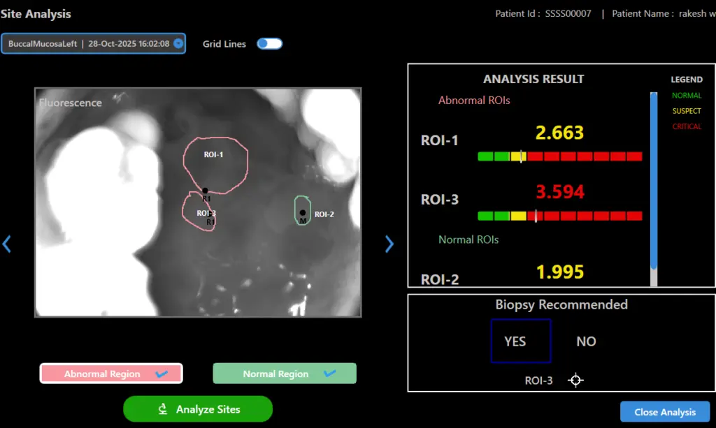
We use a patented optical engine for uniform illumination of the tissue, blocking the surface reflections and collimation of diffusely reflected light emanating from the tissue through suitable optical filters. We have built proprietary hardware and software to control sequential triggering of LEDs and synchronous image capture on a Windows environment. The captured images are processed and analyzed with the help of cloud-based machine learning algorithm to provide real-time user feedback. he system is designed to deliver consistent, high-quality imaging with minimal operator dependency, ensuring reliable performance across different clinical settings. Its advanced imaging and AI-assisted analysis support clinicians in making faster, more informed decisions while maintaining a seamless and user-friendly workflow.
- Non invasive, point of care solutions
- Accurately identifies the most malignant site for biopsy
- Reduces unnecessary repeat biopsies
- Patented and clinically validated
- Existing oral cancer screening devices rely mainly on visual examination of tissue autofluorescence or reflectance under LED illumination.
- These systems target absorption by biochemical constituents such as collagen, Protoporphyrin IX (PpIX), and NADH in tissue.
- The diagnostic outcome of current devices is highly subjective and depends heavily on the clinical expertise of the examiner.
- Clinicians often face difficulty in identifying the most malignant region for biopsy, which can result in underdiagnosis
- This limitation frequently leads to late-stage detection of oral cancer, reducing treatment effectiveness.
- To address these challenges, Sascan has developed a multispectral imaging platform that enables multimodal tissue imaging.